The Fraiya Platform: two powerful tools built for total scan confidence
FraiyaScan automates the repetitive tasks involved in prenatal scanning. FraiyaDetect adds an AI-powered layer to the secondary review process. The platform covers the complete workflow, from the moment the probe is picked up to the final report.
Available Now
20-week Anomaly Scan
Workflow automation for image capture, biometry and reporting
42% faster scans. Consistent measurements.Zero extra clicks.
A routine anomaly scan demands an extraordinary amount from the sonographer simultaneously: capturing images, measuring, tracking standard planes and staying alert for abnormalities, all on a moving fetus. FraiyaScan removes the manual burden so the sonographer can focus on the patient.
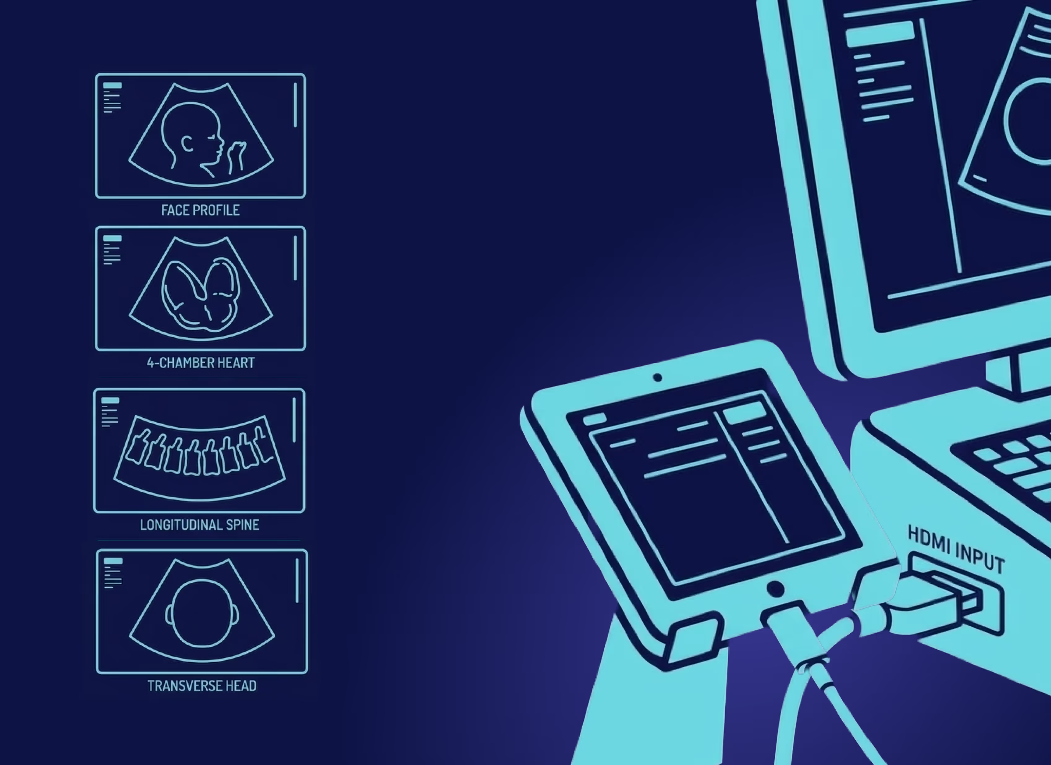
Zero-Click Image Capture
Automatically detects and saves all required standard planes from the screening protocol. No manual capture needed. No interruption.
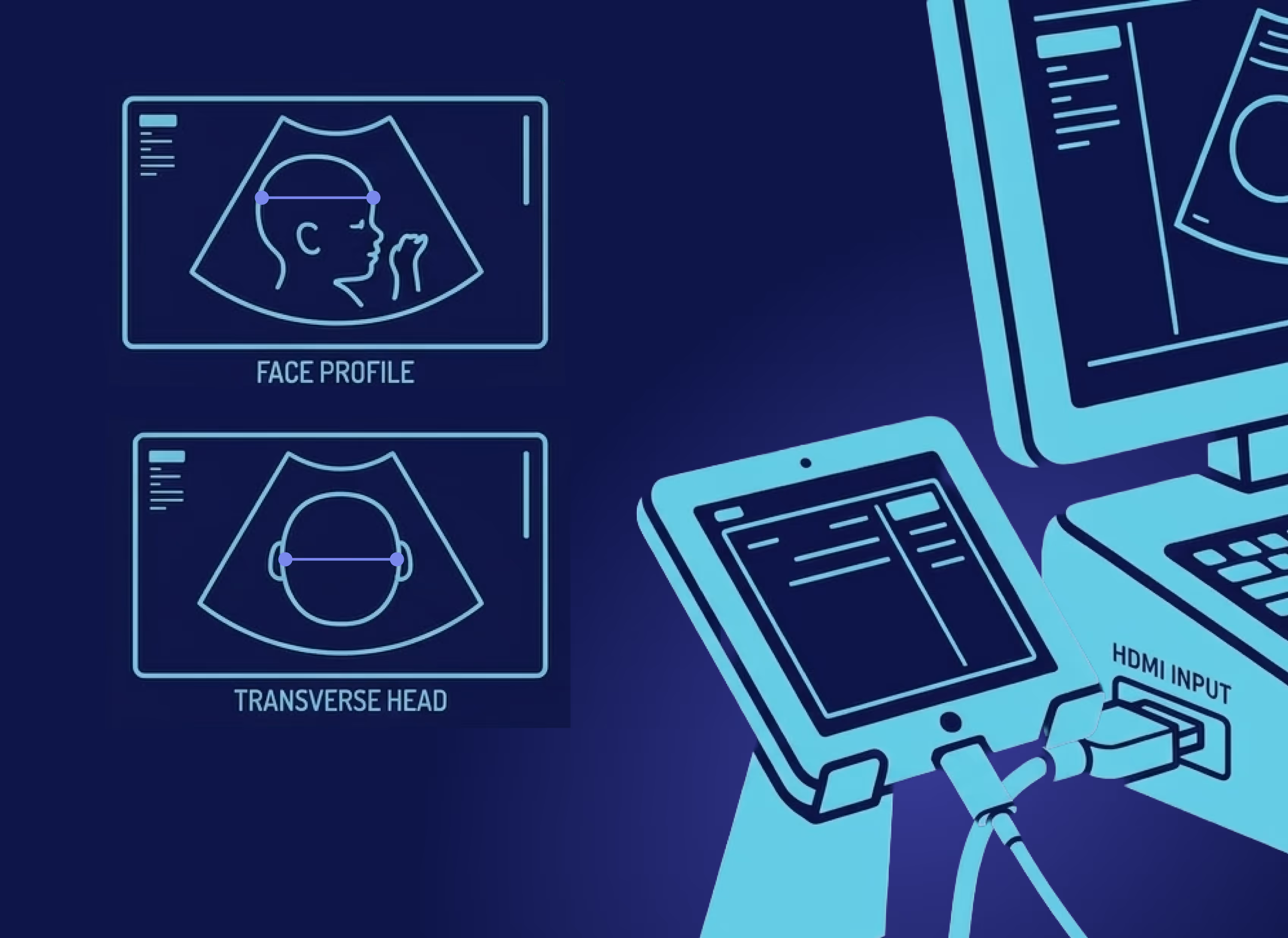
Consistent Biometry
Patented technology measures biometric parameters continuously throughout the acquisition. AI biometry is more consistent than human measurements.
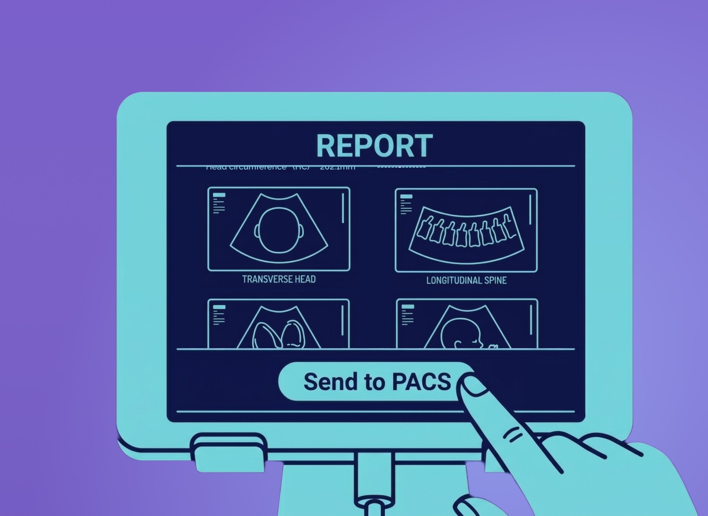
Auto Reporting
Integrates with your existing obstetric reporting packages or generates comprehensive reports. Images and measurements are sent to PACS in a single click.
FRAIYASCAN
How it Works
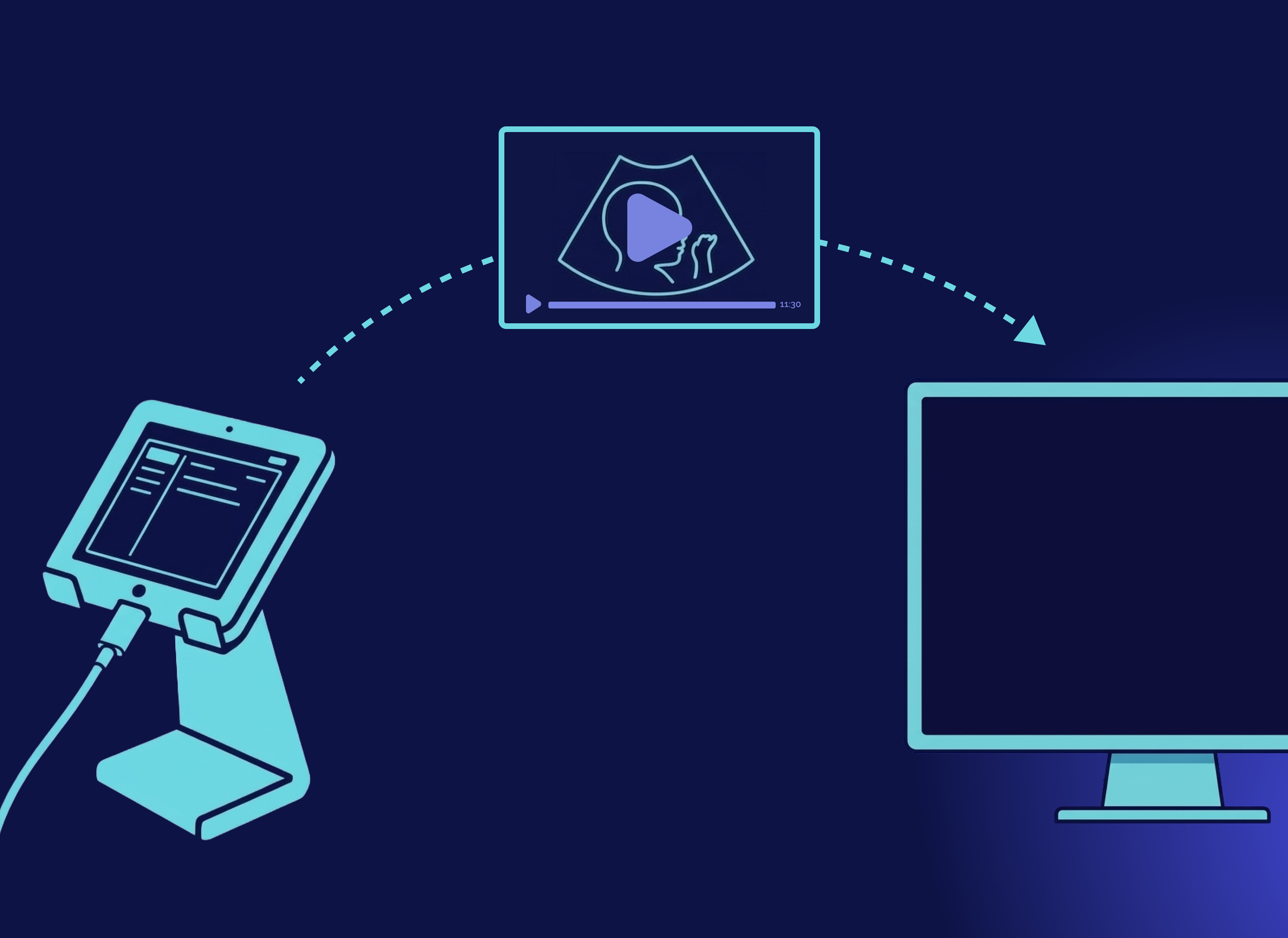
1. Connect
Connects to your existing ultrasound machines via HDMI. Department-wide licence, one installation.
2. Scan
Sonographer scans as normal. FraiyaScan captures standard planes automatically in the background.
3. Measures
Biometry taken continuously and automatically, with real-time feedback displayed on growth charts.
4. Report
Images and measurements transferred into PACS and reporting packages in one click.
Available Now
20-week Anomaly Scan
FRAIYASCAN
Outcomes
CE certified and ready for deployment across Europe. FraiyaScan was evaluated during the PROMETHEUS study, the only published randomised control trial (RCT) of AI-assisted fetal anomaly scanning in the World.1
A second opinion on every scan, powered by machine learning.
Despite advances in prenatal care, approximately half of congenital heart disease cases are still not diagnosed before birth, which has serious consequences for babies, families, and healthcare systems. Our technology uses machine learning to automatically analyse scan data and surface potential findings for clinical review.
It's not a replacement for clinical judgement. It's the safety net that reinforces it.
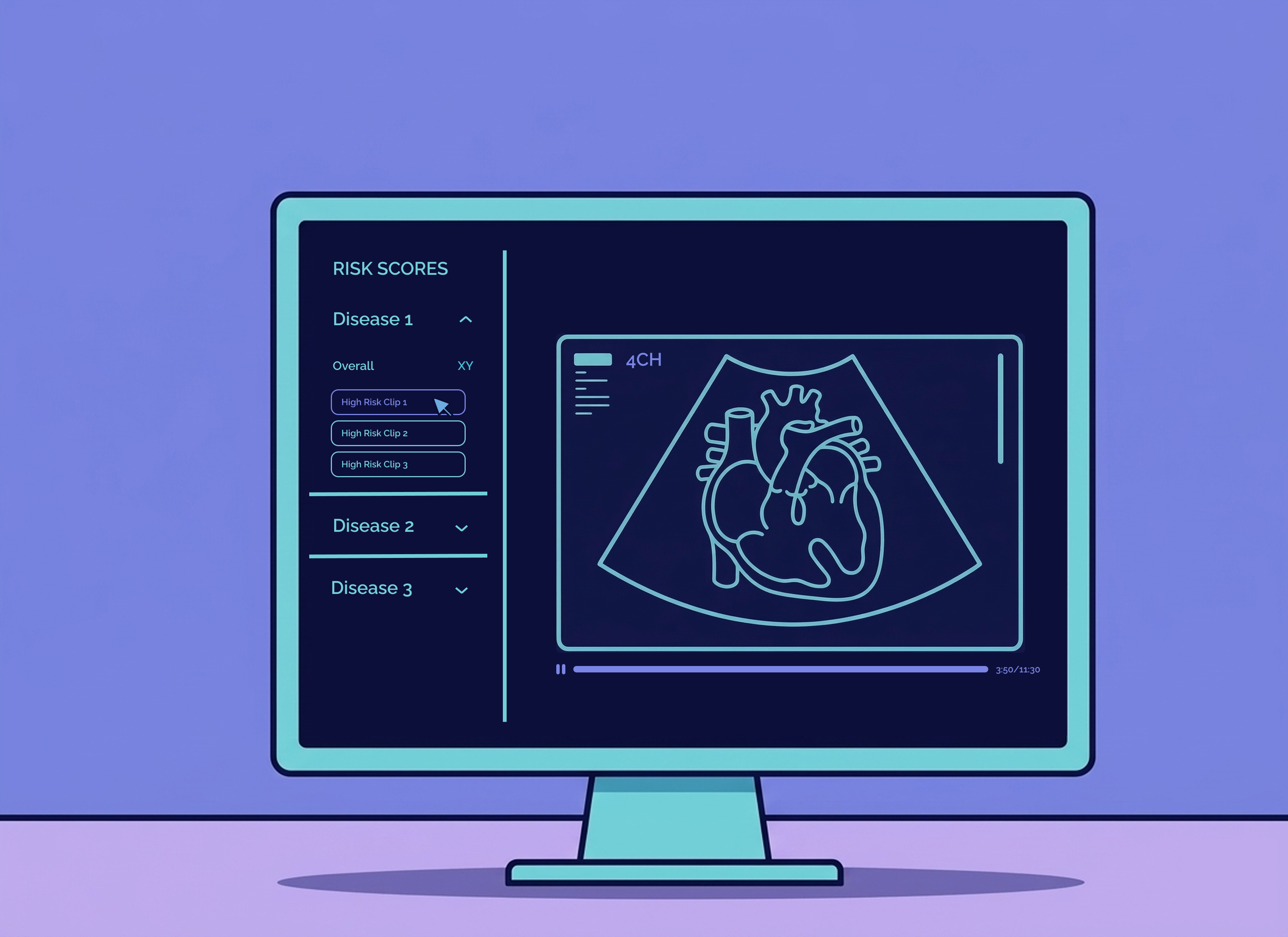
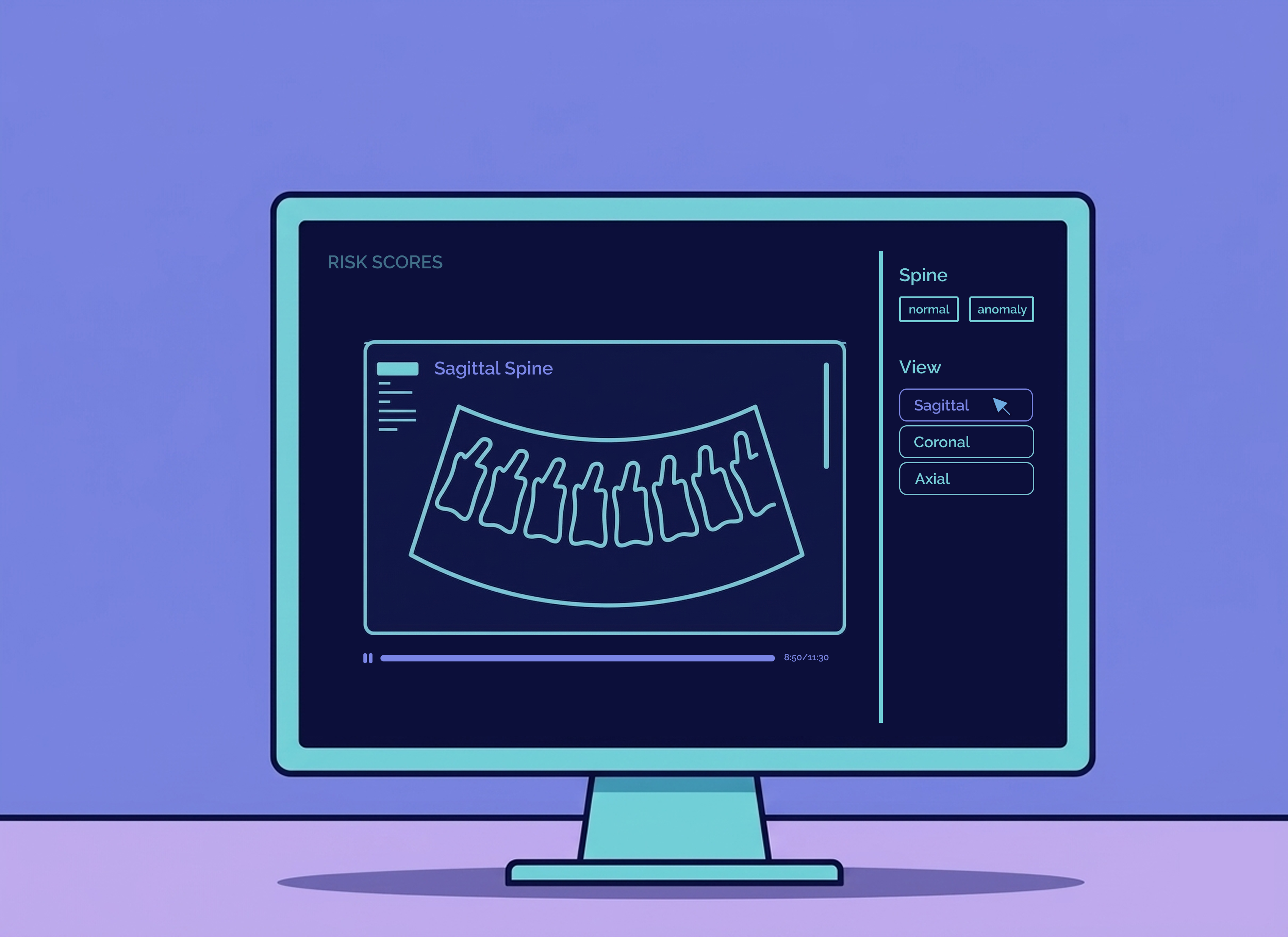
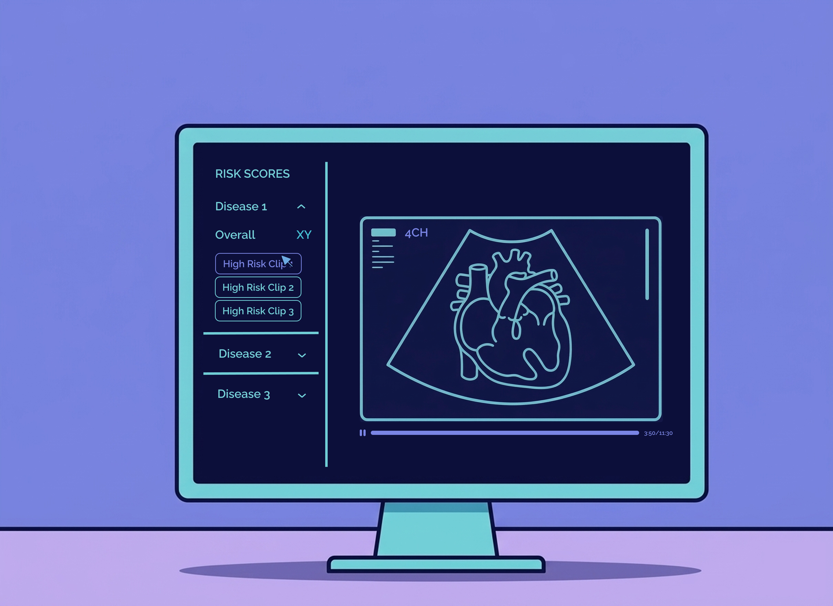
Organised scan review
Labelled clips allow rapid navigation and assessment of scans.
Cardiac risk flagging
Risk assessment of main congenital heart diseases is integrated.
Department-wide oversight
An overview dashboard provides top-level visibility of secondary review results, providing auditable records that senior clinicians can access.
FraiyaDetect
How it Works
1. Scan and Send
FraiyaScan captures and measures as normal. The whole scan is automatically pre-labelled and sent to FraiyaDetect.
2. AI analysis
ML models extract and analyse standard view clips from the scan, assigning risk scores for heart diseases.
3. Review
The secondary reviewer navigates the pre-labelled clips and has quick access to areas of concern.
4. Submit
Supporting evidence is collated and actions can be taken for clinical follow-up. Findings are logged for governance and audit.
In Development
20-week Anomaly Scan
FRAIYADetect
Outcomes
The potential of AI-assisted remote second review in fetal anomaly screening was validated by peer-reviewed research by Day et al. Our FraiyaDetect algorithms unlocked: